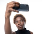
Can a single selfie really tell a computer how long someone with cancer will live?
Sounds like sci-fi. Or clickbait. Yet headlines across health media keep pointing to artificial intelligence tools trained to estimate cancer outcomes from ordinary photos of a person's face. Because the competitor URL is unavailable and has no readable text here, I can't see how that specific write-up spun the study, though the pattern is familiar. Whenever a model links a snapshot to long-term survival, people jump straight to dramatic conclusions that go way past what the data actually proves.
Here's the straight answer upfront. A photo isn't a crystal ball. When researchers train computer vision models to predict cancer survival from a facial image, the algorithm usually isn't spotting hidden tumor biology in your cheekbones. Instead, it's picking up visible clues tied to general health, aging, muscle loss, fatigue, and clinical context present in the dataset. Those clues can correlate with outcomes at a group level. They can't reliably tell you what's going to happen to an individual patient.
I've watched hype cycles around AI in medicine repeat this exact dance for years. A flashy headline promises a shortcut from a simple picture or quick scan. Clinicians read the methods section and see heavy caveats, narrow training groups, and retrospective data that still need real-world validation before anyone changes a treatment plan.
What AI Cancer Survival Predictions Actually Look For in a Face
When you hear that a tool predicts cancer survival from a selfie, it's easy to picture a camera reading microscopic cellular signals invisible to human eyes. That isn't how most of these systems work.
Oncologists have long tracked something called performance status—basically how well a patient is holding up day to day, how active they are, and how much physical toll illness or therapy has taken on their body. A person dealing with severe weight loss, cachexia, deep exhaustion, or swelling often has a tougher road ahead than someone maintaining strength and energy. Clinical scales like ECOG performance status try to capture that, though scoring can vary from doctor to doctor.
Computer vision models trained on facial photographs often end up acting as rough proxies for frailty, nutritional decline, or systemic stress. Subtle shifts in skin tone, facial volume, eye bags, or expression can reflect how worn down a body looks at a single moment. From a statistical standpoint, a group of patients who look visibly frailer on average may have different short-term or medium-term survival curves than a group that doesn't.
Correlation isn't causation. A tired look can come from poor sleep the night before, bright overhead lighting, harsh phone flash, a stressful morning in a waiting room, or medication side effects that have nothing to do with how aggressively a tumor responds to targeted therapy or immunotherapy.
Why a Single Selfie Falls Short in Real Clinical Care
Honestly, reducing a complex human health trajectory to one photo misses almost everything that matters in oncology.
Cancer isn't one disease. Lung adenocarcinoma, early-stage breast cancer, glioblastoma, and melanoma behave completely differently. Even within the same diagnosis, outcomes depend heavily on stage at diagnosis, tumor genetics and biomarkers, organ function, coexisting conditions like heart disease or diabetes, and how the cancer responds once treatment starts.
A selfie freezes one second in time. It doesn't show whether a tumor is shrinking on a CT scan, whether blood counts are rebounding after chemotherapy, or whether a patient has strong family support getting them to appointments. Treating a static image as a verdict on survival ignores the entire clinical picture.
There's also a huge gap between population statistics and personal prognosis. Suppose a model shows modest predictive accuracy across thousands of archived records. Oncologists use survival curves carefully even with gold-standard clinical data because individual patients routinely beat or defy averages. Handing someone a survival estimate generated from a photo—outside a tightly controlled research setting—can spark needless panic or false reassurance.
Hidden Biases That Skew Photo-Based Health Models
Any time an AI system learns from images and medical outcomes, dataset bias sits right around the corner. If you've spent enough time evaluating health tech, you know that flashy accuracy numbers on paper often shrink when tested on people who look different or come from different clinics.
Consider what can quietly distort a model trained on photos:
- Lighting and camera quality: Photos taken under bright clinic fluorescent lights look very different from dim home lighting or outdoor sunlight, and smartphone front cameras apply smoothing or color shifts automatically.
- Demographic representation: If a training set overrepresents certain age groups, skin tones, or care settings, the model may perform worse—or misread baseline features—in underrepresented populations.
- Data leakage and shortcuts: Sometimes models pick up background cues, grooming norms, makeup, or subtle differences in how images were captured across hospitals rather than genuine health markers.
According to guidance from the World Health Organization on AI ethics and governance for health, bias in training data and lack of real-world generalizability are central risks whenever health algorithms move from research labs toward broader use. That warning applies double when the input is something as informal and variable as a selfie.
Common Misconceptions People Have About AI Prognosis Tools
Headlines love neat narratives, so a few myths keep popping up whenever a story like this trends.
Myth 1: A selfie can replace biopsies, scans, or blood tests
No photo can tell a doctor what mutations a tumor carries or whether cancer cells have spread to lymph nodes. Tissue pathology, imaging, and lab work remain foundational because they measure biology and anatomy directly rather than guessing from surface appearance.
Myth 2: If a study is published, the tool is ready for patients
Publishing a study is an early step, not a finish line. Reliable clinical tools need prospective validation—testing the model on brand-new patients before outcomes are known—plus regulatory review and clear evidence that using the tool actually helps doctors make better decisions without harming patients. The National Cancer Institute provides clear background on how cancer prognosis is evaluated and why individual estimates carry uncertainty.
Myth 3: A scary prediction means there's nothing left to do
Even when statistical odds look tough, clinical trials, newer drug combinations, supportive care, and symptom management can change how a patient feels and how long they live. Numbers on a screen don't decide a person's grit or their next treatment option.
Where Visual AI Might Actually Help Down the Road
I'm skeptical of sensational survival claims from a snapshot, yet I don't think visual AI in oncology is pure nonsense. Used carefully, automated visual assessment could eventually support frailty screening or help flag patients who might benefit earlier from nutrition support, physical therapy, or closer monitoring.
Think about how busy clinics get. A doctor might see twenty or thirty patients in a day. If an objective, standardized tool helps flag subtle signs of physical decline that warrant a closer look—while keeping final judgment squarely with the care team—that could be genuinely practical. Still, privacy safeguards matter hugely here. Facial photos are sensitive biometric data, and patients deserve rock-solid protection over how their images are stored, shared, and used.
Practical Next Steps If You're Reading About These Studies
If you—or someone you love—are navigating cancer and stumbled across headlines about predicting survival from a selfie, take a breath and keep things grounded:
- Read past the headline: Look for what kind of cancer was studied, how many patients were included, whether the model was tested on outside data, and what the researchers actually claim it can—and cannot—do.
- Don't run photos through random apps: Avoid uploading personal or family health photos to unverified websites or consumer tools promising medical or survival predictions.
- Bring real questions to your oncology team: If you're wondering about prognosis, treatment goals, or side effects like weight loss and fatigue, ask your oncologist directly how those factors apply to your specific diagnosis.
- Focus on controllable daily habits: Talk with your care team about safe movement, protein and calorie support, sleep, and symptom relief—things that tangibly support strength and quality of life right now.
A single selfie can capture a moment on your phone. Your health story takes a whole team, real science, and much more than a picture to understand.